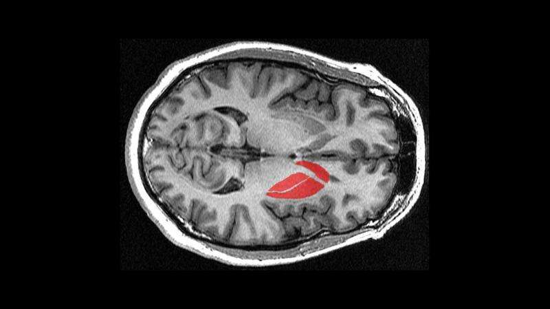
How the brain triggers action

EPFL scientists have identified specific neurons in the striatum that contribute to driving motivated behaviors like movement. The work may help in designing new ways of treating disorders like Parkinson's disease in the long term.
Perhaps the brain's most important function is to process sensory information and make behavioral decisions based on it, like moving to grasp an object. One part of the brain that does this is the "striatum", a large area in the middle of brain. Using cutting-edge techniques, neuroscientists at EPFL have now discovered how a specific type of neuron in the striatum contributes to a simple goal-directed sensorimotor behavior. The findings add a missing piece in the puzzle of the brain, contributing to our fundamental understanding of a brain area that is intimately involved in neurodegenerative disorders like Parkinson's and Huntington's disease. The work is published in Neuron.
The striatum is often described as the "coordination center" for different higher cognitive activities including motor and action planning, decision-making, motivation, reinforcement, and reward perception. To do all this, the striatum must first integrate incoming information from other parts of the brain and then respond accordingly.
The striatum thus integrates TWO sensorimotor and reward signals to guide motivated behavior. Incoming sensory and motor information reaches the striatum mostly through neurons coming from the brain's largest, and external part, the neocortex. These neurons use glutamate to transmit their signals. On the other hand, reward information comes to the striatum mostly through neurons using dopamine as their transmitter.
What does what?
The striatum contains many different types of neurons, but the distinct roles of these different types of neurons are largely unknown. To find answers, Tanya Sippy, Damien Lapray, Sylvain Crochet, and Carl Petersen at EPFL studied the striatum of mice. When one of the whiskers was briefly deflected, the animals had been trained to respond by licking a spout. This simple task allowed the researchers to monitor specific groups of neurons inside the striatum, and see which ones lit up with activity.
The first part of the experiment used a technique that can read the electrical changes occurring inside individual neurons. Here it was neurons that project from the striatum to a nearby part of the brain, the substantia nigra. This small, dark-colored area plays an important role in reward, addiction, and movement. The researchers also introduced a label into specific neurons, which allowed them to identify which type of neuron they were recording at any given time.
The experiment found that when the animal's whiskers were touched to cause them to lick the spout, certain neurons responded with what appeared to be a 'go' signal for initiating action. The neurons belong to the striatum but project from there specifically to the substantia nigra. With this finding, the researchers pinpointed a part of the brain where actions are first sparked into life.
Optogenetics
The scientists then reversed the experiment: they stimulated specific types of neurons in the striatum to see if they would cause the mice to lick. To do this, they used a technique in which Petersen's team has gained remarkable expertise over the years: optogenetics.
Optogenetics works by inserting the gene of a light-sensitive protein into live neurons. The genetically modified neurons then produce the protein, which sits on their outer membrane. There, it acts like an electrical gate, which opens when light is shone on the neuron. Ions flow into the neuron, turning it on and making it release an electric pulse. Optogenetics can also be highly targeted, stimulating specific types of neurons.
The team found that stimulating the same neurons in the striatum that had given the 'go' signal before – the ones that project to the substantia nigra – triggered the spout-licking response. In other words, the scientists circumvented the circuit, causing the same action (licking) without an external stimulus (deflecting the whickers) going first into the striatum. The optogenetic experiments added more evidence that these neurons are enough to initiate an action.
This is an important finding for fundamental neuroscience, as it identifies a specific type of neurons in the striatum where information turns into action, casting light on a core function of the brain. But there is long-term potential too, as the study highlights a means for possible treatments of diseases that affect the action initiation and motor control, such as Parkinson's and Huntington's.
More information: Tanya Sippy et al. Cell-Type-Specific Sensorimotor Processing in Striatal Projection Neurons during Goal-Directed Behavior, Neuron (2015). DOI: 10.1016/j.neuron.2015.08.039


