Fruit fly muscles with a hypertrophic cardiomyopathy mutation don't relax properly

Using fruit flies, Johns Hopkins researchers have figured out why a particular inherited human heart condition that is almost always due to genetic mutations causes the heart to enlarge, thicken and fail. They found that one such mutation interferes with heart muscle's ability to relax after contracting, and prevents the heart from fully filling with blood and pumping it out.
More specifically, the researchers say, the molecular machinery of the cells' "skeleton," designed to shorten muscle, is more prone to locking in place and remaining partially contracted.
Because the mutation is in a protein conserved throughout evolutionary history, the researchers say, the findings, reported online Sept. 12 in Cell Reports, could help guide approaches to treating human hypertrophic cardiomyopathy.
Hypertrophic cardiomyopathy, marked by a thickening of the human heart's lower chambers, which are comprised of heart muscle tissue, occurs in an estimated one in 500 men and women, often without symptoms. It can lead to an irregular heartbeat, heart pumping failure and sudden death in people under 35.
The particular mutation the researchers used to genetically engineer the fruit flies for their study is known in humans as ACTC A295S. ACTC is a protein called alpha-cardiac actin that helps form contractile elements of the heart muscle cells' cytoskeleton. The mutation or alteration substitutes the 295th amino acid in the protein's backbone—an alanine—for a serine, and was discovered in 1999 in 13 family members with hypertrophic cardiomyopathy by scientists in Denmark. More than 1,500 mutations in nine genes have been identified as causes of hypertrophic cardiomyopathy.
"The protein affected by this particular hypertrophic cardiomyopathy mutation is extremely well-conserved across the animal kingdom, and the region where the mutation is located is 100 percent identical in a thousand different animal sequences, meaning that it is highly likely that the mutation will have the same effect on human muscle tissue as it does in fruit flies," says Anthony Cammarato, Ph.D., assistant professor of medicine at the Johns Hopkins University School of Medicine. "Because this protein is so essential to muscle function, and in higher organisms there are additional versions of it to compensate for the mutant form in animal cells, scientists have struggled to duplicate the precise disease alteration in a model organism to show how the mutation causes disease, until now."
To see how the fly version of ACTC A295S works on heart muscle, the researchers used genetic tools to either turn on an extra normal copy or the mutant form of the actin protein in the fruit fly heart.
The fruit fly heart is a single tube of tissue that fills with hemolymph, or "fly blood," after relaxing, and then pumps it along. The researchers videoed the hearts pumping and then compared the hearts with the normal and the mutant actin. They noticed that the hearts with the mutant actin were shorter in diameter or skinnier than the hearts with the normal copy of actin. In one-week-old flies, the pumping capacity was about 85 nanoliters per minute in the flies with mutant actin in the hearts compared to 125 nanoliters per minute in the hearts with the normal actin.
To find out why the hearts with the mutant form of actin were shorter in diameter than the ones with normal actin, the investigators soaked the hearts in a chemical that sucks up all the calcium from the heart, which normally causes hearts to completely relax and the diameter to expand.
Both healthy and mutant hearts increased in diameter by about 2.5 percent, but the mutant hearts were still shorter than normal. Next, the researchers added the drug blebbistatin to the calcium-removing chemical and soaked the hearts. Chains of actin form filaments in cells, and a second set of filaments composed of the protein myosin grab onto actin and pull the muscle filaments over one another so they overlap, shortening the cell and causing muscle contraction. Blebbistatin prevents myosin from binding to the actin and contracting the muscle cells. After treatment with blebbistatin, the hearts with the normal actin relaxed another 2 percent, but the hearts with the mutant actin relaxed another 8 percent.
"The myosin doesn't completely release from the actin in the hearts expressing the hypertrophic cardiomyopathy mutation, which prevents the heart from entirely relaxing or fully filling," says Cammarato.
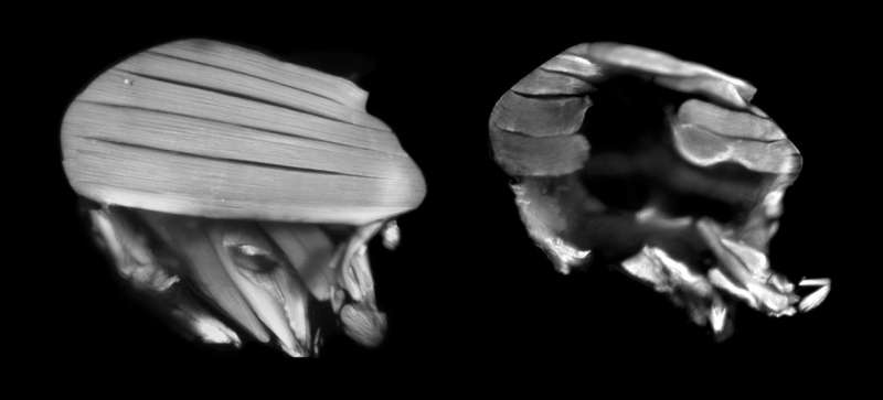
In another set of experiments, the researchers took advantage of the fact that the muscles that power flight are larger than the flies' hearts, making it easier to study muscle mutations and their biomechanical effects. In fruit flies that were bred to lack actin in their flight muscles, the researchers added back a normal copy of actin or a hypertrophic cardiomyopathy mutation version into the fly's genome. They then looked at the flight muscles in both sets of flies. In the flies with the normal copy of actin the flight muscles looked normal, but in the flies with the mutant actin the muscles looked shredded, which the researchers say was from excessive force in the muscles because they couldn't properly relax.
In muscle tissue, the protein tropomyosin lies along the actin filaments and prevents myosin from attaching and contracting the muscle. Tropomyosin is the "gatekeeper" of activating muscle contraction. Once an electrical signal comes through, calcium is released into the cell and tropomyosin moves out of the way, allowing myosin to temporarily bind to the actin filaments and contract the muscle.
To figure out why myosin doesn't seem to completely release from the actin filaments in the flight and heart muscles of flies with the hypertrophic cardiomyopathy mutation, the researchers used computer simulations to investigate the molecular changes behind the overly contracted muscles.
The researchers calculated the energy of the electrostatic interaction between tropomyosin and normal actin compared to hypertrophic cardiomyopathy actin. They found that tropomyosin has a 60 percent smaller area to move over the actin filaments when interacting with the mutant actin compared to the normal actin, and that tropomyosin's position on the mutant actin has likely moved away from a location that prevents myosin from binding.
"We think that this altered positioning and restricted motion of tropomyosin prevents it from properly bumping myosin off the actin filaments when it's time for the muscle to relax, which is what is keeping the muscles contracted, and in humans may ultimately trigger disease" says Cammarato.
More information: Meera C. Viswanathan et al. Distortion of the Actin A-Triad Results in Contractile Disinhibition and Cardiomyopathy, Cell Reports (2017). DOI: 10.1016/j.celrep.2017.08.070


