Near-atomic map of parathyroid hormone complex points toward new therapies for osteoporosis

An international team of scientists has mapped a molecular complex that could aid in the development of better medications with fewer side effects for osteoporosis and cancer.
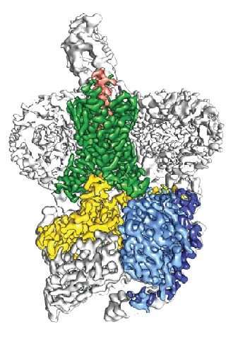
The near-atomic resolution images depict parathyroid hormone receptor-1 (PTH1R), a molecule that conveys signals to and from cells, interacting with two key messengers—a molecule that mimics parathyroid hormone, one of the most important regulators of calcium levels in the body, and a stimulatory G protein, a molecule that mediates bone turnover.
The findings, published today in Science, give researchers a better blueprint for designing drugs for osteoporosis and other conditions such as chachexia, which causes severe weakness and weight loss that can be fatal in cancer patients.
Globally, more than 200 million people have osteoporosis and even more have low bone density. In the coming years, public health experts expect these numbers to rise, fueled in part by an aging population. They also fear an increase in osteoporosis-related fractures due to fewer people taking current medications out of concern over rare side effects.
"The understanding of how all of these molecules fit together has been a missing piece of the puzzle since the discovery of parathyroid hormone 80 years ago," said H. Eric Xu, Ph.D., a professor at Van Andel Research Institute (VARI) and co-corresponding author of the study. "It's a big step forward that we hope will one day help people around the world."
PTH1R is a molecular communication conduit between cells and their environments that fosters development of the bones, skin and cartilage, and regulates levels of calcium in the blood.
To do this, it interacts with molecular messengers such as the parathyroid hormone, which ensures the blood stream has the appropriate amount of calcium to maintain healthy function.
However, too much parathyroid hormone can wreak havoc on the body, spiking the amount of calcium in the blood to dangerous levels, promoting the formation of kidney stones and leaching calcium from bones, which can cause devastating fractures. Too little bogs down metabolism, and contributes to fatigue, weight gain, depression and a host of other issues.
Today's findings also provide insight into G protein-coupled receptors (GPCRs), a family of signaling molecules to which PTH1R belongs. Taken together, GPCRs are targeted by nearly 30 percent of medications currently on the market.
GPCRs are notoriously difficult to visualize using traditional X-ray crystallography methods; to date, only about 40 out of more than 800 total GPCRs have had their structures determined. To visualize today's structure, the team used a groundbreaking technique called cryo-electron microscopy (cryo-EM), which is capable of imaging molecules in unprecedented clarity and can more easily image molecules like GPCRs that are embedded in the cell membrane.
More information: "Structure and dynamics of the active human parathyroid hormone receptor-1" Science (2019). science.sciencemag.org/cgi/doi … 1126/science.aav7942


