Experimental noninvasive tool monitors effectiveness of stem cell transplantation
Stem-cell based therapies to strengthen the heart muscle and treat other diseases are beginning to show promise in human clinical trials. However, other than observing clinical outcomes, lack of a repeatable, time-sensitive and noninvasive tool to assess the effectiveness of the transplanted cells within the target organ has slowed progress in the stem cell field.
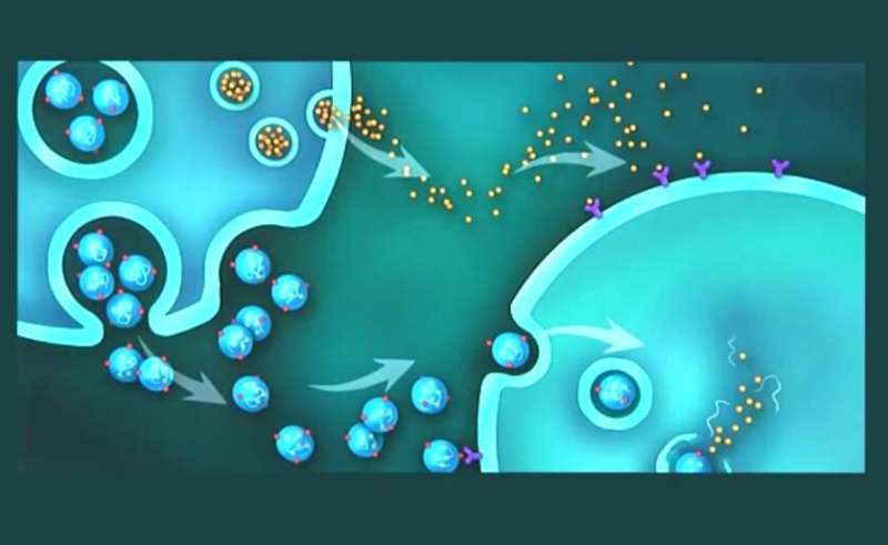
Researchers from the University of Maryland School of Medicine (UMSOM), the University of Pennsylvania and Emory University theorized that a blood test could be used to track the efficacy of transplanted stem cells. They aimed to achieve their goal by analyzing tiny cellular components called exosomes, secreted from the transplanted stem cells into the recipient blood. They tested their theory in rodent models of heart attack, or myocardial infarction, after transplanting two types of human cardiac stem cells and monitoring their circulating exosomes. The researchers found circulating exosomes delivered cell components to the target heart muscle cells, resulting in cardiac repair. Results are published in the journal Science Translational Medicine.
"Exosomes contain the signals of the cells they're derived from—proteins as well as nucleic acids and micro ribonucleic acids (miRNAs) - which affect receptor cells and remodel or regenerate the organ we're targeting," said study co-senior author Sunjay Kaushal, Ph.D., MD, Professor of Surgery at UMSOM and Director of Pediatric Cardiac Surgery at the University of Maryland Children's Hospital. "We now have a tool to determine whether stem cell therapy will be efficacious for an individual patient, not only for the heart but for any organ that received stem cell therapy."
Through the blood test, which the researchers call a "liquid biopsy," the researchers monitored human cardiosphere-derived cells (CDCs) and cardiac progenitor cells (CPCs) transplanted into rat hearts following myocardial infarction. Blood plasma concentrations of the exosomes were compared seven days after transplant.
After purifying the CDC/CPC-derived exosomes, the researchers found the exosomes contained miRNAs associated with heart muscle recovery. Further, they found CPCs and CDCs produced in culture differed in contents from exosomes produced by transplanted cells in the living organism.
"Our study should be considered the first stepping stone in understanding what stem cells do, but an important point is that the cells we identified as responding changed their gene expression, behavior and secretions," said co-lead author Sudhish Sharma, Ph.D., UMSOM Assistant Professor of Surgery. "By using these biomarkers, we can understand the mechanism and extent of recovery."
More information: P. Saha el al., "Circulating exosomes derived from transplanted progenitor cells aid the functional recovery of ischemic myocardium," Science Translational Medicine (2019). stm.sciencemag.org/lookup/doi/ … scitranslmed.aau1168


