New bone imaging technique could lead to improved osteoporosis treatment

More than 2.3 million Canadians are affected by osteoporosis, resulting in billions of dollars in economic burden and incalculable suffering.
A research team from the College of Medicine at the University of Saskatchewan has developed a new approach to imaging that detects changes in bone tissue far more quickly than bone densitometry scans, the method currently used in health care. While the study was done using a rabbit model, the results could lead to improved drug treatment in humans with osteoporosis.
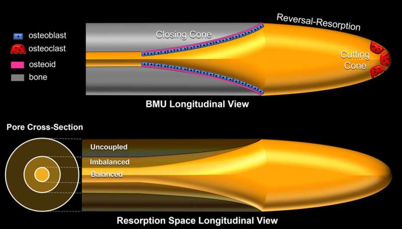
Using the BMIT beamline of the Canadian Light Source at the University of Saskatchewan, Dr. David Cooper and colleagues were able to see the incredibly tiny pores inside cortical bone, the dense outer surface of bone that accounts for the majority of bone mass. These pores change over time, showing how bone tissue is continuously removed and replaced.
The researchers stimulated this bone turnover using parathyroid hormone, then tracked the changes in the pores of the cortical bone in as little as 14 days.
"In humans, the pores we were looking at are about the width of a few hairs—a quarter of a millimeter—and in rabbits they're about half that size," said Cooper, whose latest breakthrough builds on a decade's worth of work in this area. "Using micro-computed tomography (micro-CT) we were, for the first time, able to see the shapes of these pores and actually track them over time."
Study lead Dr. Kim Harrison said this research would not have been possible using conventional X-ray techniques. "This uses refractive qualities between soft and hard tissues which highlights these pores within the bone and makes it easier to image and track the changes," she said.
"This really is the establishment of a fundamentally new way of looking at bone turnover," said Cooper. "Nobody has ever been able to do this before in terms of tracking the pores."
Current osteoporosis diagnostic testing, done by bone density scans, is "a very blunt instrument," Cooper added. "It might take years for changes to be detectable. We're detecting these things over weeks in animals. It could have a rapid impact on how current drugs used in the treatment of osteoporosis are deployed."
The research was published in the Journal of Bone and Mineral Research.
More information: Kim D Harrison et al, Direct Assessment of Rabbit Cortical Bone Basic Multicellular Unit Longitudinal Erosion Rate: A 4D Synchrotron‐Based Approach, Journal of Bone and Mineral Research (2022). DOI: 10.1002/jbmr.4700


