This article has been reviewed according to Science X's editorial process and policies. Editors have highlighted the following attributes while ensuring the content's credibility:
fact-checked
proofread
Advanced MRI technique unleashes new approach for diagnosing diabetic kidney disease
 9.4-T vertical standard-bore super-conducting magnet, (B) The 1H and 23 Na surface coil. Credit: Kidney360 (2023). DOI: 10.34067/KID.0000000000000072")
The gold standard test for predicting the onset of diabetic kidney disease is the albuminuria test. However, detecting albuminuria alone has limited sensitivity and specificity in end-stage renal failure with a decreased estimated glomerular filtration rate. This is supported by several reports, which state that about half of the type 2 diabetes patients who developed kidney dysfunction showed no preceding albuminuria.
In this study, the authors propose the possibility of diagnosing tubular abnormalities in diabetic kidney disease at an early stage and connecting them to treatment by combining the evaluation of sodium concentration using 23Na MRI with the measurement of urinary albumin, commonly used in diabetic testing.
23Na MRI, which images 23Na instead of the clinically applied 1H in existing MRI, visualizes metabolic changes in sodium. Although there have been previous reports of 23Na MRI studies, capturing sodium concentrations equivalent to physiological saline and visualizing them clearly in MRI has been challenging because the amount of sodium in vivo is much smaller than hydrogen, resulting in extremely small signal values. This study optimized the newly developed 23Na MRI to image the kidneys of mice, enabling detailed and clear imaging.
The kidney is involved in the excretion and reabsorption of sodium. It is equipped with mechanisms such as the countercurrent multiplication system, which allows efficient water reabsorption, and the formation of an osmotic gradient by sodium ions from the cortex to the medulla. Although many studies have been conducted on channels and transporters involved in renal sodium metabolism, the changes in kidney's overall amount and distribution of sodium need to be explored further. The advanced 23Na MRI technique can noninvasively evaluate the distribution of sodium throughout the kidney and assess changes in the countercurrent multiplication system.
-
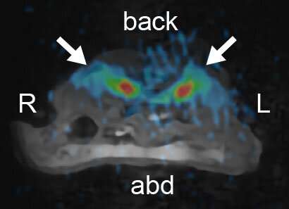
23Na MR images merged with 1H MR images (C57BL/6JJcl mice, 6 weeks old, male). This image shows the transverse plane through the abdomen. Arrows indicate the kidneys. MR, magnetic resonance; 23Na, sodium; 1H, proton; L, left; R, right; abd, abdomen Credit: Kidney360 DOI: 10.34067/KID.0000000000000072 -
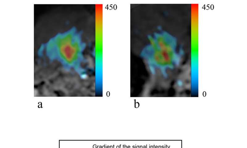
(A, B) 23Na MR image merged with 1H MR image of the right kidney in an m+/m+ mouse (A) and a db/db mouse (B). (C) The signal intensity gradients from the cortex to the medulla in m+/m+ and db/db mice. The signal intensity of the medullary region in the kidney was lower in db/db mice than in m+/m+ mice. The sodium concentration gradients from the medulla to the cortex of m+/m+ and db/db mice were 2.28±0.21 and 1.64±0.36, respectively (P < 0.01). Credit: Kidney360 DOI: 10.34067/KID.0000000000000072
This study used six-week-old diabetic model mice (db/db mice) at an early stage when no tissue damage was observed in the kidneys and imaged the kidneys using a 9.4 Tesla vertical magnet MRI device optimized by the research group. Compared to control mice, the formation of the countercurrent multiplication system in the kidney was weakened, as revealed by the 23Na-visualization images. The success of this study is attributed to the research group's invention of the advanced MRI device and their unique imaging techniques.
23Na MRI can reveal metabolic changes in renal sodium and may enable a better understanding of the pathophysiology of not only diabetic kidney disease but also dehydration and some forms of hypertension related to sodium metabolism. In the future, the researchers plan to investigate whether these findings can help diagnose other diseases. Moreover, the successful detailed visualization of the mouse kidneys suggests the possibility of expanding the target organs. Accumulating these findings may open new avenues for the clinical application of 23Na MRI.
The article "Sodium magnetic resonance imaging shows impairment of the countercurrent multiplication system in diabetic mice kidney" was published in the journal Kidney360.
More information: Yusuke Nakagawa et al, Sodium Magnetic Resonance Imaging Shows Impairment of the Counter-current Multiplication System in Diabetic Mice Kidney, Kidney360 (2023). DOI: 10.34067/KID.0000000000000072


