This article has been reviewed according to Science X's editorial process and policies. Editors have highlighted the following attributes while ensuring the content's credibility:
fact-checked
peer-reviewed publication
trusted source
proofread
Non-invasive technology maps brain activity to investigate behavior changes in neurological disease

A research team led by Cleveland Clinic and Oregon Health and Science University (OHSU) has developed a new method for mapping how the parts of the brain "speak" to each other, critical to understanding behavior changes in patients with neurological disease.
Diseases like Alzheimer's disease change how patients communicate and act, affecting their relationships and well-being. Cleveland Clinic's Hod Dana, Ph.D., is collaborating with Jacob Raber, Ph.D., an OHSU behavioral neuroscientist, on mapping out the electrical paths that connect and coordinate the parts of the brain needed to complete different tasks.
"Effects on behavior and personality in Alzheimer's disease and related disorders are caused by changes in brain function," Dr. Dana says. "If we can understand exactly how the changes occur, we may figure out how to slow down the process or to stop it. Recording brain activity patterns that underlie behavioral changes is the first step to bridging the gap."
Decision-making, forming a memory or completing a task all involve brainwaves, signaling pathways that use cells called neurons. To study how brainwaves influence behavior and decision making, researchers observe as neurons turn "on" and "off" across the organ in different situations.
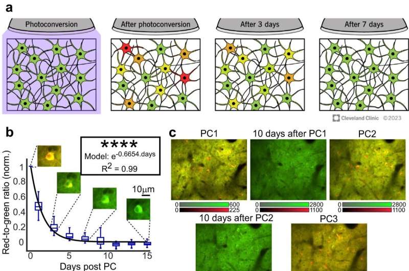
Current technologies are unable to map the whole brain while still identifying the single cells. CaMPARI images can be captured during behavior, highlighting neurons that are active as red and inactive neurons as green. After the test is completed, the red and green markers remain bright for several days. This allows researchers to capture a series of images to track the brain's activity by mapping where the red appears within the brain.
The team have published results in Nature Communications on using a calcium sensor system called CaMPARI (Calcium-modulated photoactivatable ratiometric integrator) to map brain activity in preclinical models while completing cognitive tasks. Drs. Dana and Raber plan to use CaMPARI in preclinical work to see how Alzheimer's-related genes affect the way our neurons signal through our brains in learning and memory.
Drs. Dana and Raber say they hope to take what they learn from their results to develop tests and interventions that can improve the quality of life for patients, providing better treatment options.
"We now have the capability to study the relationship between brain activation and cognitive performance at an unprecedented level," says Dr. Raber. "These are the first steps in developing strategies to reverse those changes and improve cognitive performance in those affected by neurological conditions. The future of behavioral and cognitive neuroscience looks bright."
More information: Aniruddha Das et al, Large-scale recording of neuronal activity in freely-moving mice at cellular resolution, Nature Communications (2023). DOI: 10.1038/s41467-023-42083-y


