This article has been reviewed according to Science X's editorial process and policies. Editors have highlighted the following attributes while ensuring the content's credibility:
fact-checked
peer-reviewed publication
trusted source
proofread
3D bioprinting advances research on respiratory viruses

The COVID-19 pandemic has profoundly impacted our lives, claiming nearly 7.1 million lives globally. Scientists and medical professionals have been working tirelessly to understand the virus, its transmission pathways, and effective treatments.
The urgency of developing vaccines and treatments has never been greater, prompting calls for Korea to expedite its drug development efforts to match those of developed countries. Recently, a team of Korean researchers has made a breakthrough that will greatly enhance the efficiency of respiratory disease research, garnering attention.
The research team, consisting of Professor Sungjune Jung and Ph.D. student Yunji Lee from the Department of Materials Science and Engineering at POSTECH, in collaboration with Dr. Meehyein Kim and Dr. Myoung Kyu Lee from the Infectious Diseases Therapeutic Research Center of the Korea Research Institute of Chemical Technology (KRICT), has successfully created artificial lungs.
These artificial lungs are designed to study infections and test drugs for respiratory diseases including COVID-19. Their research has been published in Biomaterials.
On average, developing a new drug takes 10 to 15 years and costs over 1 trillion won. This lengthy and expensive process is largely due to existing research platforms, such as 2D cell cultures and animal experiments, which fail to accurately replicate the in vivo environment. To reduce development time and costs and to increase the success rate, models that closely mimic the human body are essential.

In this study, researchers from POSTECH and the KRICT created a "3D artificial lung" using three-dimensional (3D) bioprinting technology. This advanced technology uses cells and biomaterials to produce lifelike tissues and organs, reducing the need for animal testing in fields such as regenerative medicine and drug discovery.
The "3D artificial lung" created by the researchers consists of three layers—vascular endothelium, extracellular matrix, and epithelium— just like the human respiratory tract. This model closely resembles the structure and function of the human lung including cell-cell junctions and mucus secretion. It also contains high levels of proteins (ACE2, TMPRSS2) that serve as entry points for the COVID-19 virus at the epithelial layer, making it susceptible to infection even at very low doses.
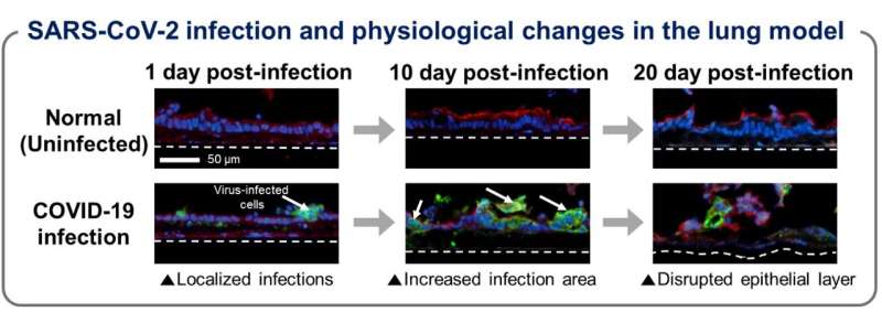
Unlike conventional 2D cell culture models which destroy cells within five days of infection, the researchers' model lasted 21 days. This duration is sufficient to observe infection-induced cell lesions and barrier degradation, enabling the team to identify changes in gene expression affecting the infection pathway, viral proliferation, and host immune response, consistent with real-world COVID-19 patient data.
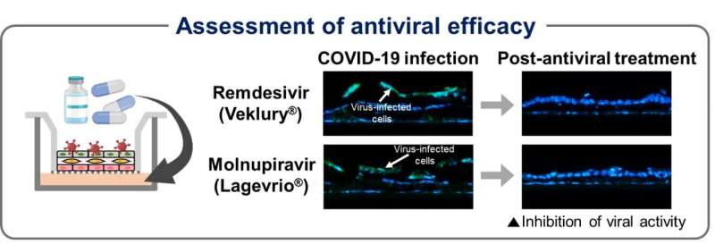
Moreover, the team perfectly recreated the pathways by which COVID-19 drugs (Remdesivir and Molnupiravir) reach the infected epithelial layer and inhibit virus replication. Unlike conventional 2D cell culture methods where drugs are directly administered to the epithelial cells, this study evaluated the drug efficacy after they crossed the tissue barrier through the artificial lung tissue. This approach allowed the researchers to accurately verify the effectiveness of the treatments, determine appropriate dosages, and identify potential side effects.

Professor Jung remarked, "Academics are warning that respiratory viruses similar to COVID-19 are likely to emerge in the next decade." He added, "This research will not only dramatically shorten the drug development process but also aid in developing therapeutic drugs for COVID-19 and other respiratory diseases."
Dr. Kim from the KRICT stated, "To prepare for a pandemic like COVID-19, we need to enhance early efficacy evaluation systems by using 3D cell and tissue culture technology that considers clinical relevance. Additionally, it is essential to promote research on therapeutic development for human-infectious viruses with this technology."
More information: Yunji Lee et al, 3D-printed airway model as a platform for SARS-CoV-2 infection and antiviral drug testing, Biomaterials (2024). DOI: 10.1016/j.biomaterials.2024.122689


