October 21, 2011 report
Cerebellar neurons needed to navigate in the dark
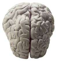
(Medical Xpress) -- A new study by scientists in France has revealed that the cerebellum region of the brain plays an important role in the ability to navigate when visual cues are absent, and is the first study to show this kind of influence of the cerebellum on the hippocampus, which was already known to be involved in the kind of mental mapping needed for navigation.
Previous studies have demonstrated that three kinds of cell in the hippocampus allow animals to navigate. There are neurons representing direction, which fire when the animal is facing a specific direction, neurons for place (place cells), which fire only when the animal is in a specific place, and other neurons known as grid cells, which fire regularly as an animal moves, tracking the animal’s movements, and enabling connections to be made between place, direction and spatial movements.
Until the present study the communication networks between the cerebellum and hippocampus in spatial movement tasks were unknown. The researchers, led by Laure Rondi-Reig of the Université Pierre et Marie Curie in Paris, said their study demonstrates a two-way communication between these regions of the brain.
The study used genetically engineered mice that had defective expression of the cerebellar enzyme protein kinase C, or PKC, which is known to be involved in the neural processing of cues concerning body movements, balance, and depth perception. Using transgenic mice lacking an effective enzyme allowed the team to compare their abilities with normal mice and so determine what connections, if any, the cerebellar neurons had with those in the hippocampus.
One experiment that compared these abilities used a water maze, in which six genetically engineered mice and five controls were trained to swim in the maze to a platform in the center. The mice performed equally well when the lights were turned on, but when the experiment was repeated in the dark, the transgenic mice had trouble finding their way around the maze.
In the darkness the hippocampus place cells fire at a lower rate in all the mice, which, Dr Rondi-Reig said meant they had to rely on the cerebellar neurons involved in self-motion cues. In the PKC-deficient mice these neurons did not work as effectively, and so they were less sure of the direction in which they should move.
The results of the experiments therefore suggest that spatial mind maps produced in the hippocampus depend on mechanisms in the cerebellum that require the PKC enzyme.
More information: Cerebellum Shapes Hippocampal Spatial Code, Science 21 October 2011: Vol. 334 no. 6054 pp. 385-389. DOI:10.1126/science.1207403
Abstract
Spatial representation is an active process that requires complex multimodal integration from a large interacting network of cortical and subcortical structures. We sought to determine the role of cerebellar protein kinase C (PKC)–dependent plasticity in spatial navigation by recording the activity of hippocampal place cells in transgenic L7PKCI mice with selective disruption of PKC-dependent plasticity at parallel fiber–Purkinje cell synapses. Place cell properties were exclusively impaired when L7PKCI mice had to rely on self-motion cues. The behavioral consequence of such a deficit is evidenced here by selectively impaired navigation capabilities during a path integration task. Together, these results suggest that cerebellar PKC-dependent mechanisms are involved in processing self-motion signals essential to the shaping of hippocampal spatial representation.
© 2011 MedicalXpress.com


