Getting the measure of MRI
(Medical Xpress) -- A method for imaging the brain that has largely been confined to neuroscience labs may now find its place as a proper tool for medical diagnosis.
Oxford University scientists have come up with a new approach that turns functional magnetic resonance imaging (fMRI) from something that produces pictures of changes in brain activity into a full numerical measure of how the brain is working.
Doctors may be able to use this new MRI approach to provide a lot more clinically useful information about patients coming in with strokes, brain injuries or a variety of other conditions.
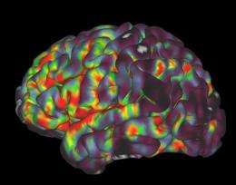
Functional MRI is a tremendously successful research method for imaging the brain. The pictures it produces of the working brain are now pretty familiar, with different regions of the brain ‘lighting up’ while those being scanned do different tasks, and it has taught us a lot about the organisation of the working brain in health and disease.
However, the technique only captures relative changes in the MRI signal. And what is more, the MRI signal reflects a complex mix of different physiological processes going on in the brain, such as changes in blood flow and brain cells burning up the oxygen they get from the blood. That is: fMRI is very much an indirect indicator of brain activity, not a pure measurement technique able to put a value on specific brain processes.
"MRI is great for localizing which areas of the brain are activated during different stimuli and so helping us to understand how the brain works as a whole," explains Dr. Daniel Bulte of Oxford's Centre for Functional Magnetic Imaging of the Brain (FMRIB), who led the work. "However the images we produce are just that, pictures. They are not measurements."
The scientists at FMRIB have countered this by introducing a new approach to MRI scanning. They report their work in the journal NeuroImage.
Patients lie in the MRI scanner and breathe air through a mask or nose tubes. By simply varying the proportion of carbon dioxide and oxygen the patients breathe through the mask, the scientists show it is possible to use the MRI signal to measure blood flow, blood volume, oxygen use and brain metabolism across the whole brain.
"By making some slight changes to the air breathed by a patient in the scanner we can produce beautiful images of brain physiology that actually correspond to real measurements," says Daniel. "During the scan the subject would spend short periods breathing a little extra oxygen than normal and short periods breathing a little extra carbon dioxide. The subject would not notice either of these as the changes are quite subtle compared to normal air."
The measurements are comparable to those obtained with another more complex medical technique called Oxygen-15 positron emission tomography, or 15O-PET. However, the MRI approach would have a number of advantages and – importantly – wouldn’t expose patients to the radioactive labels used in PET.
"The problem with 15O-PET is that it is very, very expensive, there are very few places in the world that can do it, the scans take a long time, and it requires giving the patient a significant dose of radiation," says Daniel. "Our MRI method takes less than 20 minutes to perform, could be run on any modern clinical MRI scanner, is very cheap and uses no drugs or injections, and exposes the patient to no ionising radiation."
Daniel adds that it could be a real boon in hospitals: "At the moment most stroke patients get a CT scan when they arrive at the hospital, and very few get an MRI. The main reason for this is that MRI is much more expensive than CT, and the imaging techniques currently available do not provide enough diagnostic information to be sufficiently useful for most patients. Thus the diagnoses and treatments are arrived upon with very little detailed information about what is actually going on in the patient's brain.
"A scan such as this one could potentially be used to provide emergency room staff with much more information about a variety of different diseases and injuries, improving the outcomes for the patients, and saving time and money. We hope to start trialling the scans with patients with a range of different diseases later this year."


