Surgeons use woman's own tissue to rebuild ear lost to cancer
(Medical Xpress)—In a series of a half-dozen operations spanning 20 months, surgeons at Johns Hopkins have successfully reconstructed the entire ear and part of the skull of a 42-year-old woman from Bel Air, Md.
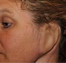
The patient's left ear had been almost entirely removed to spare her life, threatened by an aggressive skin cancer that had also spread to nearby parts of her temporal skull, parotid salivary gland and inner left ear canal, which were also surgically removed.
The painstaking series of operations—begun in January 2011 and ending with ear-contouring and shaping surgery in September 2012—is believed to be one of the most complicated ear reconstructions ever performed at Johns Hopkins, according to the facial plastic and reconstructive surgeons involved in the case. Piecing together more than a dozen bits of bone, cartilage, skin and arteries, they rebuilt the patient's ear and inserted a bone-anchored hearing aid to restore her hearing. All living tissues were taken from spare or renewable parts of her own body to minimize the risk of rejection by her immune system.
"It's my skin, my bone, and the most realistic surgical replacement to what my ear was before my cancer," says Sherrie Walter, a working mother of two whose ordeal began in 2008 with the scab that "just didn't heal."
The retail sales manager says she is still somewhat shocked at her diagnosis of basal cell carcinoma because she does not think of herself as a typical skin cancer patient. Skin cancer doesn't run in her family, and although she has occasionally taken trips to the beach, she says she is no sun worshipper.
"I didn't know how aggressive basal cell carcinoma could be," says Walter, whose initial diagnosis in early 2008 led to a session of intense radiation therapy and regular skin biopsies to monitor any cancer growth.
In October 2010, she discovered some blood in her left ear and learned that her cancer had returned. That Christmas, surgeons at Johns Hopkins removed her ear, as well as other surrounding head, neck, gland, lymph and skull tissues to which her tumor had spread.
"When my doctors told me reconstruction was possible, I thought it was too good to be true; it sounded like science fiction," says Walter, who adds, "Just learning that reconstructing my ear was doable gave me sufficient physical and emotional strength, as well as the confidence I needed to go through with the surgeries."
Lead surgeon Patrick Byrne, M.D., says Walter's reconstructive options were few, as her cancer surgery had removed the surrounding and underlying skull bone structure needed to support more traditional options, such as a re-attachable, plastic prosthetic ear.
Further complicating Walter's ear reconstruction was that the surrounding, stretchable facial and neck skin normally used for the outside skin covering was also not available. It, too, had been lost during her initial surgery. Also lost was a key portion of her skull bone needed for an implantable, prosthetic ear option.
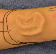
Byrne, who has performed hundreds of ear and nose reconstructions, says facial and neck skin is ideal because it matches well to the outside ear and can be stretched and regrown. In Walter's case, however, he says his team "had no choice" but to look at other options for an outside skin covering. They chose forearm skin as the next best match.
After removing pieces of rib cartilage to assemble the new ear, Byrne and his team carved and sutured the cartilage together in a semicircular pattern precisely measured to match Walter's other ear.
The newly assembled—still skinless—ear was then implanted under Walter's forearm skin for four months so that the skin, with its own nourishing blood vessels, could stretch and grow around the new ear. Walter also had reconstructive surgery performed on her skull base to reinforce the new ear.
Aiding Byrne in the January 2012 surgery was an intra-operative laser, with a low-enough frequency to penetrate skin without burning it, yet high enough to visualize the ultra-thin blood vessels inside the ear. With it, Byrne was able to assess if the new ear had sufficient blood flowing through it and where. He then affixed three of the largest of the hard-to-find blood vessels—one artery and two veins—in the newly attached ear to matching blood vessels in the head.
Byrne, an associate professor in otolaryngology-head and neck surgery at the Johns Hopkins University School of Medicine, praises Walter's commitment to her recovery.
"Patients must have the physical and emotional courage, and the patience, to deal with these exhausting procedures, and to recover and re-energize so they can proceed from one surgery to the next," says Byrne, who is also director of facial plastic and reconstructive surgery at Johns Hopkins.
More information:
For additional information, go to:
www.hopkinsmedicine.org/facial … s/patrick_byrne.html
www.hopkinsmedicine.org/news/m … g_his_own_body_parts


