Study reveals information about the genetic architecture of brain's grey matter

An international research team studying the structure and organization of the brain has found that different genetic factors may affect the thickness of different parts of the cortex of the brain.
The findings of this basic neuroscience study provide clues to better understanding the complex structure of the human brain. Ultimately, knowledge of genetic factors that underlie brain structure may help to identify individuals at risk for neuropsychiatric disorders, such as autism, schizophrenia or dementia. However, further research is necessary and the road to preventing or treating these conditions based on this work remains a long one.
The team was led by researchers at the University of California, San Diego, and included scientists from Virginia Commonwealth University, Boston University, Harvard Medical School and Massachusetts General Hospital, the University of Helsinki in Finland and the Veterans Affairs San Diego Healthcare System.
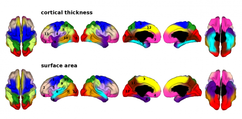
In the study, published online this week in the Proceedings of the National Academy of Sciences Online Early Edition, the team used MRI brain scan data collected from more than 200 pairs of twins between the ages of 55 and 65 and created a map based on genetic correlations between measures of thickness at different places on the cortex.

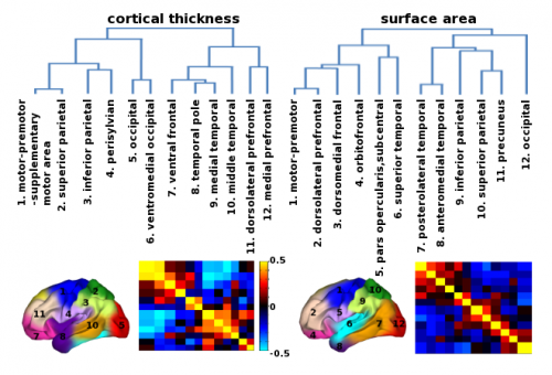
Using software developed by Michael Neale, Ph.D., professor of psychiatry and human genetics in the VCU School of Medicine, the team drew a genetic correlation map based on cortical thickness at thousands of points on the surface of the brain. These correlations were then analyzed to identify regions where the same genetic factors seem to have been operating. Twelve such regions in each hemisphere were identified, similar to an earlier study of measures of surface area.
"Our team has mapped genetic factors that influence the thickness of the cortex of the human brain," said Neale who was a study contributor and co-author.
"Knowledge of the genetic organization of brain structures may guide the identification of risk factors for psychiatric disorders," he said.
According to Neale, individuals differ in the thickness of these regions, and a twin study can help differentiate genetic from environmental factors that cause these differences at any one location. Twin studies also can estimate the degree to which the same versus different genetic factors affect two different characteristics.
Traditionally, maps of the human brain have been drawn using one of two types of information. The first is anatomical, such as the wrinkles on the surface, or cortex, of the brain. A second type of map, which may be called functional, is drawn from knowledge of how different parts of the brain are associated with particular functions. For example, Wernicke's area on the left side of the brain is associated with the understanding of language.

The research builds on work published last year in Science by the same research team. That article reported on the initial development of the new software tool to study and explain how the brain works. It was considered the first map of the surface of the brain based on the basis of genetic information.
Next steps for this research will include correlating measures of these regions with outcomes, such as change in cognitive abilities since age 20, or lifetime cigarette smoking.
For nearly 30 years, Neale, an internationally known expert in statistical methodology, has developed and applied statistical models in genetic studies, primarily of twins and their relatives, with the goal of better understanding the brain and behavior.


