To better understand the brain, look at the bigger picture

Researchers have learned a lot about the human brain through functional magnetic resonance imaging (fMRI), a technique that can yield insight into brain function. But typical fMRI methods may be missing key information and providing only part of the picture, Yale researchers say.
In a new study, they evaluated various approaches and found that zooming out and taking a wider field of view captures additional relevant information that a narrow focus leaves out, offering greater understanding of neural interplay.
Further, these more encompassing results may help address neuroimaging's reproducibility problem, in which some findings presented in studies cannot be reproduced by other researchers.
The findings were published Aug. 4 in Proceedings of the National Academy of Sciences.
Studies employing fMRI typically focus on small areas of the brain. As one example of this approach, researchers look for brain regions that become more "active" when a particular activity is performed, homing in on small areas with the strongest activation. But a growing body of evidence shows that brain processes, and complex processes in particular, aren't limited to small parts of the brain.
"The brain is a network. It's complex," said Dustin Scheinost, an associate professor of radiology and biomedical imaging and senior author of the study. Oversimplifying, he said, leads to inaccurate conclusions.
"For more sophisticated cognitive processes, it's unlikely that many areas of the brain are wholly uninvolved," added Stephanie Noble, a postdoctoral associate in Scheinost's lab at Yale School of Medicine and lead author of the study. Focusing on small areas leaves out other regions that may be involved in the behavior or process under study, which can affect the direction of future research as well.
"You develop this incorrect picture of what's actually happening in the brain," she said.
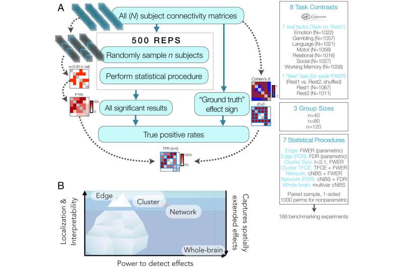
For the study, researchers assessed how well fMRI analyses across a range of scales were able to detect effects, or changes in fMRI signals as participants perform different activities, revealing which parts of the brain are engaged. They used data from the Human Connectome Project, which has collected brain scans of individuals as they performed different tasks related to complex processes like emotion, language, and social interactions. The research team looked for effects in very small parts of the brain network—such as connections between just two areas—and also at clusters of connections, widespread networks, and whole brains.
They found that the larger the scale, the better they were able to detect effects. This ability to detect effects is known as "power."
"We get better power with these broader-scale methods," said Noble.
At the smallest scales, researchers were only able to detect about 10% of effects. But at the network level, they could detect over 80% of them.
The trade-off for the extra power was that the broader views didn't relay information that is as spatially exact as that of smaller scale analyses. For instance, at the smallest scale, researchers could confidently say that the effects they observed were occurring throughout the small area. At the network level, however, they could only say that the effects were occurring across much of the network, not pinpoint where in the network exactly.
The goal, Noble says, is to balance the benefits and disadvantages of the various methods.
"Would you rather be very confident about a small portion of the relevant information—in other words, have a very clear picture of just the tip of the iceberg?" she said. "Or would you rather have a really big picture of the whole iceberg that's maybe a little bit blurry but gives you a sense of the complexity and wide spatial scale of where things are occurring in the brain?"
For other researchers, this approach is simple to implement, and Noble said she looks forward to seeing how other scientists use it.
She notes that the fields of psychology and neuroscience, neuroimaging included, have faced a reproducibility problem. And low power in fMRI analyses contribute to it: low-power studies only uncover small portions of the story, which can be seen as contradictory rather than parts of a whole. Increasing fMRI power, as she and her colleagues did here by increasing the scale of their analyses, could be one way to address reproducibility challenges by exposing how seemingly contradictory results may in fact be harmonious
"Moving up the food chain, so to speak, going from very low level up to more complex networks buys you a lot more power," said Scheinost. "This is one of the tools we can use to help the reproducibility problem."
And scientists shouldn't throw the baby out with the bathwater, Noble said. There's plenty of good work being done to improve methods and boost rigor, and fMRI is still a valuable tool, she said: "I think assessing power, rigor, and reproducibility is healthy for any field. Especially one that deals with the complexity of living beings and mental processes."
Noble is now developing a "power calculator" for fMRI, to help others design studies in a way that achieves a desired level of power.
More information: Stephanie Noble et al, Improving power in functional magnetic resonance imaging by moving beyond cluster-level inference, Proceedings of the National Academy of Sciences (2022). DOI: 10.1073/pnas.2203020119


