This article has been reviewed according to Science X's editorial process and policies. Editors have highlighted the following attributes while ensuring the content's credibility:
fact-checked
peer-reviewed publication
trusted source
proofread
Researchers develop method to measure protein expression in neurons

Northwestern Medicine investigators have developed a method to measure protein expression in an individual neuron, a discovery that will enable scientists to study how this process goes awry in disease, according to a study published in Molecular Psychiatry.
Neurons in the brain communicate primarily through chemical signals and disruption in neuronal communication can lead to disorders such as autism, Parkinson's and Alzheimer's disease. Previously, scientists had no way of measuring protein expression within a single type of neuron, which limited how closely they could study dysfunctional neurons.
"We were motivated to make these new tools to actually study how synaptic proteins are changed and how these processes could contribute to the functional deficits seen in specific neurological disorders," said Jeffrey Savas, Ph.D., assistant professor in the Ken and Ruth Davee Department of Neurology's Division of Behavioral Neurology and senior author of the study.
In the study, Savas and his collaborators developed synaptic probes, which consisted of a virus activated by a protein present in a specific type of neuron. Once inside a mouse and activated, the virus would release a protein capable of "tagging" other proteins expressed by the neuron, allowing investigators to pinpoint, monitor, and quantify the proteins expressed in a particular type of neuron.
First, the investigators studied two types of neurons in the striatum, an area of the brain involved in decision-making functions, such as motor control, emotion and habit formation. According to the study, they found that protein expression was similar in both direct and indirect pathway spiny projection neurons (dSPNs and iSPNs), two of the major neuron types in the striatum.
"The surprise was that while the protein identities were not dissimilar, the expression of the isoforms was," Savas said. "Isoforms are like cousins of proteins that are related, but they might be shorter or longer but are still encoded from the same gene."
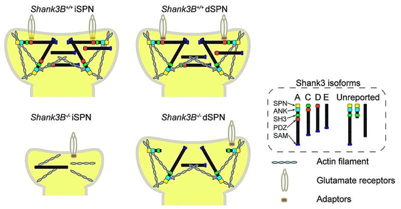
In particular, the isoforms produced by the gene SHANK3 in neurons were starkly different, Savas said. SHANK3 has long been thought to play a key role in autism spectrum disorders, but confounded scientists studying it because mutations in the gene led to markedly different symptoms across individuals.
By tracking neuronal protein expression with their new probes in Shank3b knockout mice, the investigators discovered that several SHANK3 isoforms were expressed by dSPNs but were undetectable in iSPNs, which resulted in hampered neuronal communication in mice.
Notably, these dSPNs appeared healthier than the iSPNs, a difference that contributed to an imbalance in synaptic transmission within the basal ganglia. Remarkably, by reintroducing Shank3b isoforms in the knockout mice, investigators were able to rescue many of the synaptic deficits, according to the study.
The findings may also explain why people with autism caused by SHANK3 mutations can present with a wide range of symptoms, Savas said.
"This highlights a new aspect of molecular specificity in the mammalian brain," Savas said. "Human patients who have mutations in the SHANK3 gene phenotypically look very different, but the mutations are in the same gene.
"The most compelling possibility by which that happens is that those different mutations affect isoform-specific expression of SHANK3 protein isoforms in a cell-type-specific manner. The manifestation of that mutation impacts different parts of the brain and then those different parts of the brain are responsible for different phenotypes associated with autism."
"Currently, we are applying these probes, to additional synapses, cell types, and different mouse models to see if we can elucidate similar mechanisms in the context of other neurological disorders," Savas said.
More information: Yi-Zhi Wang et al, Neuron type-specific proteomics reveals distinct Shank3 proteoforms in iSPNs and dSPNs lead to striatal synaptopathy in Shank3B–/– mice, Molecular Psychiatry (2024). DOI: 10.1038/s41380-024-02493-w


