This article has been reviewed according to Science X's editorial process and policies. Editors have highlighted the following attributes while ensuring the content's credibility:
fact-checked
peer-reviewed publication
trusted source
proofread
Scaffolding sensors detect early evidence of organ transplant rejections in mice

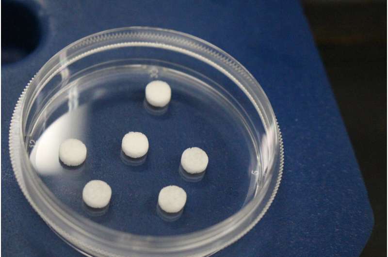
A new microporous scaffold functions as a minimally-invasive surveillance method to identify rejection prior to graft injury in a mouse model, according to a study published in Science Advances.
These sensors are a first step towards developing a tool which could provide physicians with vital early information about the possibility of organ rejection in transplant patients.
Solid organ transplantation is associated with aggressive immunosuppression to prevent graft rejection. However, over suppression can increase the risk of neoplasia and opportunistic infections, while insufficient suppression can lead to graft injury.
Traditionally, biopsies of a transplanted organ are performed to assess the efficacy of immunosuppression. However, these invasive biopsies have significant variability and are a lagging indicator of rejection. To identify rejection prior to graft injury, a University of Michigan research team used a microporous scaffold that functions as a minimally-invasive surveillance method.
Following heart or skin transplantation in mice, niche implants accumulate innate and adaptive immune cells, and gene expression analyses identify biomarkers of acute cellular allograft rejection (ACAR) prior to clinical signs of graft injury.
Initial studies were performed with adoptive transfer of T cells with mismatched allografts that enabled a focus on T cell-mediated rejection, with subsequent validation studies in wild-type animals. The scaffold niche permits frequent sampling of the cells, and the gene biomarker panel distinguishes mice rejecting allogeneic grafts from those with healthy grafts.
"Research on monitoring immune responses has been exciting given the rise in immunotherapies. This detection of an undesired immune response has substantial medical opportunities, because you often do not learn about an undesired response until an organ begins to lose its function," said Lonnie Shea, the Steven A. Goldstein Collegiate Professor of Biomedical Engineering at U-M and corresponding author on the study.
The new process starts by implanting a porous scaffold under the skin, where tissue develops within the pores. The tissue that develops becomes vascularized. The net effect is that blood vessels are running through this space, with immune cells circulating through those vessels.
The material elicits a foreign-body response that leads to recruitment of immune cells. Importantly, these cells exhibit a phenotype representative of tissues rather than those in circulation, allowing researchers to monitor tissue responses over time.
"When the immune system becomes activated in the context of transplant rejection, you can see activated immune cells at the implant," said Shea.
The ability to assess immune responses within tissues can be a powerful tool for researchers to learn about the immune system. By sequencing the cells' transcriptomes, they can detect possible organ rejection with a minimally invasive biopsy instead of performing a biopsy of the transplanted organ that would have a greater risk profile.
"The survival of solid organ transplants has been heralded as one of the hallmarks of modern medicine, yet we often overlook the aggressive therapies needed post-transplant to maintain healthy grafts," said Russell Urie, a postdoctoral research fellow of biomedical engineering at U-M.
"These implant sensors can identify very early processes in rejection, which is the first step towards a tool for personalizing post-transplant care and minimizing the invasive procedures and devastating side effects that transplant recipients must currently tolerate," said Urie.
"This will be of particular impact for pediatric and young adult organ graft recipients as they must undergo multiple decades of treatment and biopsies and even retransplantation."
More information: Russell R. Urie et al, Biomarkers from subcutaneous engineered tissues predict acute rejection of organ allografts, Science Advances (2024). DOI: 10.1126/sciadv.adk6178


