Functional MRI provides support in operations on the brain
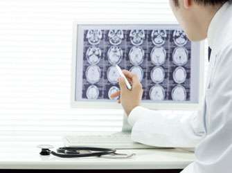
Researchers at the MedUni Vienna have proved in a so far unique multicenter study that clinical functional magnetic resonance tomography (fMRI), in the area in which the MedUni Vienna has a leading role internationally, is a safe method in brain surgery. With the aid of fMRI imaging can pinpoint to the millimetre where critical nerve fibres (e.g. vital for speech or hand function) lie and which have to be avoided – in operations on brain tumours for example.
"With the assistance of functional magnetic resonance tomography we are, if you like, drawing a red line for the surgeon so he knows where not to make an incision so as to avoid damage," says Roland Beisteiner from the University Department of Neurology at the MedUni Vienna. The neurologist and president of the Austrian Society for fMRI was playing a part in the development of fMRI as early as 1992, initiating its development in Austria. Since then this method has been developed and implemented at the University Department of Neurology and the High Field MRI Center of Excellence.
Now Beisteiner's team have been able for the first time to demonstrate in a current paper in the top journal "Radiology" that functional magnetic resonance tomography provides diagnostic certainty in operations on the brain – no matter what the equipment is (whether a 7Tesla magnetic resonance tomograph as in Vienna or even only a 1.5Tesla), no matter in which location and also irrespective of who is operating it. The Medical Universities in Innsbruck and Salzburg, the Heinrich Heine University of Düsseldorf and the Stiftungsklinikum Koblenz (Koblenz Hospital Foundation) also took part in the study.
The "Imaging and Cognition Biology" Research Cluster of the MedUni and Vienna University
Likewise, with the help of functional magnetic resonance tomography, the teams of Beisteiner and Tecumseh Fitch (Faculty of Life Sciences of the University of Vienna) are investigating in a joint research cluster belonging to the MedUni Vienna and the University of Vienna whether the structural and syntactic processing of music takes place in similar areas of the brain as does the processing of speech. Says Beisteiner: "It is never exactly the same area of the brain; however, brain activities can overlap when talking or playing an instrument."
The main focus of the research cluster is to determine precisely the common areas of the brain involved and to develop new treatments by activating them. These could perhaps then be used on people suffering from aphasia, which is a loss of language as the result of brain damage mostly to the left half of the brain.
According to Beisteiner there have been some astonishing results: "People, who could no longer speak because of their aphasia, have been able to sing the words they have learned to the matching tune." From this one can conclude that it would seem to make sense to also practise music skills during speech therapy.
The "Imaging and Cognition Biology" research cluster is one of six joint clusters at the MedUni Vienna with the University of Vienna, which were set up in 2011. Further information: forschungscluster.meduniwien.ac.at/.
More information: Wurnig, M. et al. Variability of Clinical Functional MR Imaging Results: A Multicenter Study. March 22, 2013, doi: 10.1148/radiol.13121357


