This article has been reviewed according to Science X's editorial process and policies. Editors have highlighted the following attributes while ensuring the content's credibility:
fact-checked
peer-reviewed publication
trusted source
proofread
'Mini-brain' study reveals possible key link to autism spectrum disorder

Using human "mini-brain" models known as organoids, Mayo Clinic and Yale University scientists have discovered that the roots of autism spectrum disorder may be associated with an imbalance of specific neurons that play a critical role in how the brain communicates and functions. The specific cells are known as excitatory cortical neurons.
The new study is published in Nature Neuroscience.
Findings
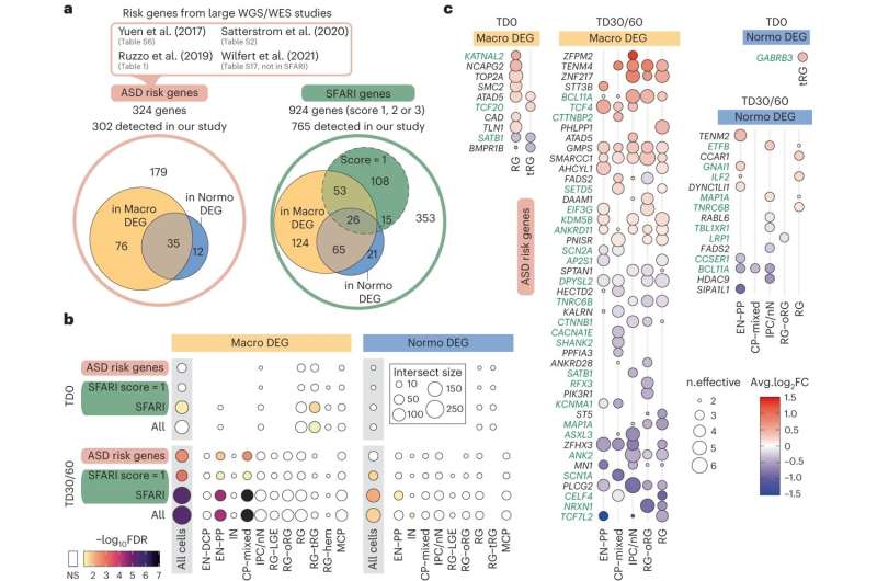
The team found an abnormal imbalance of excitatory neurons in the forebrain of people with the disorder, depending on their head size.
"This organoid technology allowed us to recreate the brain development alteration that happened in the patients when they were in the uterus, which is believed to be the time when autism spectrum disorder originates," says Alexej Abyzov, Ph.D., a genomic researcher in the Department of Quantitative Health Sciences at the Mayo Clinic Center for Individualized Medicine. Dr. Abyzov is a senior author of the study.
What is autism spectrum disorder
Autism spectrum disorder is a neurological condition that affects the way people perceive and interact with others, leading to challenges in social communication and behavior. The term "spectrum" emphasizes the broad range of symptoms and severity, and includes autism, Asperger's syndrome, childhood disintegrative disorder and an unspecified form of pervasive developmental disorder.
Nearly 1 in 36 children in the U.S. has been identified with autism spectrum disorder, according to estimates from the Centers for Disease Control's Autism and Developmental Disabilities Monitoring Network.
More about 'mini-brains'
For the study, the scientists first created miniature 3D brain-like models, called organoids. The pea-sized clusters of cells began as skin cells from people with autism spectrum disorder. The skin cells were placed in a culture dish and "reprogrammed" back into a stem-cell-like state, called induced pluripotent stem cells. These so-called master cells can be coaxed to develop into any cell in the body, including brain cells.
Next, the scientists used a special technology called single-cell RNA sequencing to study the gene expression patterns of individual brain cells. In all, they examined 664,272 brain cells at three different stages of brain development.
The scientists also discovered that the neuron imbalance stemmed from changes in the activity of certain genes known as "transcription factors," which play a crucial role in directing the development of cells during the initial stages of brain formation.
Building evidence
This study builds on 13 years of published studies on autism spectrum disorder by Dr. Abyzov and his collaborators, including Flora Vaccarino, M.D., a neuroscientist at Yale University. In one pioneering study, they showed molecular differences in organoids between people with autism and those without and implicated the deregulation of a specific transcription factor called FOXG1 as an underlying cause of the disorder.
"Autism is mostly a genetic disease. Our goal is to be able to determine the risk of autism spectrum disorder and possibly prevent it in an unborn child using prenatal genetic testing. However, this would require detailed knowledge of how brain regulation gets derailed during development. There are many aspects in which organoids could help in this direction," says Dr. Abyzov.
More information: Jourdon, A. et al. Modeling idiopathic autism in forebrain organoids reveals an imbalance of excitatory cortical neuron subtypes during early neurogenesis. Nature Neuroscience (2023). DOI: 10.1038/s41593-023-01399-0 www.nature.com/articles/s41593-023-01399-0


