May 16, 2024 report
This article has been reviewed according to Science X's editorial process and policies. Editors have highlighted the following attributes while ensuring the content's credibility:
fact-checked
peer-reviewed publication
trusted source
proofread
Miniaturized optical coherence tomography imaging probe takes pictures inside cerebral arteries

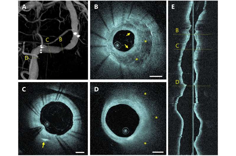
A large international team of micro-engineers, medical technologists, and neurosurgeons, has designed, built and tested a new type of probe that can be used to take pictures from inside arteries in the brain.
In their paper published in the journal Science Translational Medicine, the group describes how the probe was designed and created and how well it has performed during initial testing.
When patients develop medical problems in their brains, such as clots, aneurysms or hardened arteries, the tools available to doctors to diagnose them are limited to imaging technology that takes pictures of veins and arteries from an outside-of-the-brain view. Such images are then used as maps to direct catheter-like devices through veins and arteries into and through parts of the brain to make repairs.
The problem with such an approach is that the imagery used is not always clear, or precise. It also does not allow the surgeon to see what is happening inside of the vein or artery as it is being repaired, resulting in semi-blind procedures.
In this new study, the research team has created a camera-carrying probe that is small enough to fit inside a catheter, which allows for the capture of near-real-time imagery from inside veins and arteries in the brain.
The new probe is based on optical coherence tomography—a type of imaging technology that has been used by eye and heart surgeons to treat patients. It generates images by processing backscattering of near-infrared light. Until now, such devices have been too bulky and stiff for use inside the brain.
To overcome this problem, the research team replaced the components with smaller parts, such as a fiber-optic cable as thin as a human hair. They also used a modified type of glass to forge a distal lens that makes up the head of the probe that allows for bending.
The resulting probe is mostly hollow and has a wormlike appearance. It also spins at 250 times per second to help it move easily through veins and arteries. The camera takes pictures at a rate proportional to need. The entire probe fits easily inside a catheter, which facilitates its placement and movement inside the arteries and veins of the brain—and also its removal.

After testing in animals, the probe has been moved to clinical trials at two sites, one in Canada, the other in Argentina. To date, 32 patients have been treated using the new probe. The team reports that thus far, they have found it to be safe, well-tolerated and successful in all cases. They conclude by suggesting their new probe is ready for general use.
More information: Vitor M. Pereira et al, Volumetric microscopy of cerebral arteries with a miniaturized optical coherence tomography imaging probe, Science Translational Medicine (2024). DOI: 10.1126/scitranslmed.adl4497
© 2024 Science X Network


