Algorithm provides real-time neurosurgery link

Neuro-navigation technology is set to change the face of neurosurgery, with a simple computer algorithm able to convert pre-operative brain images into a real-time picture of the position of healthy and diseased tissues during surgery.
Deputy Head of UWA's School of Mechanical and Chemical Engineering Winthrop Professor and lead researcher Karol Miller says "for us as simple mechanical engineers, the problem of brain deformation and shift during surgery is a mechanical problem – it is, scientifically speaking, a problem in solid mechanics".
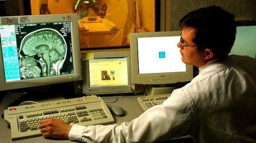
W/Prof Miller, who is also the director of the university's Intelligent Systems for Medicine Laboratory, says neurosurgeons in the majority of hospitals around the world use pre-operative MRI images to guide surgery.
He says this method does not account for the deforming of tissue including brain shift which occurs during the opening of the skull, surgical manipulation and removal of diseased tissue.
"The neurosurgeon makes a guess about how the brain shifted after opening the skull," he says
"The surgical target and critical healthy areas of the brain may move by 10mm or more which means that the surgery is usually performed very conservatively because if they cut too much there will be serious deficits."
W/Prof Miller and his team have developed computer algorithms that use the geometry from pre-operative MRI and the real-time position of the exposed surface of the brain to give accurate images throughout surgery.
"We are now able compute the deformations of the brain and display deformed or "warped" pre-operative images to the surgeon so that the image corresponds to the current intra-operative position of the tumour and critical healthy areas that need to be avoided in the approach," he says.
"We modestly say that our method is not inferior to the use of intra-operative MRI, an approach that requires multimillion dollar equipment, and have demonstrated that there is a 98 per cent probability that at least 25 per cent of patients, in which only pre-operative MRIs are used, would benefit."
The group now plan to trial their technique at Sir Charles Gardiner Hospital, whereby surgeons using the traditional pre-operative MRI will have the added ability to view the image produced by the algorithm.
The surgeons would then be able to give feedback on whether this new information would change their surgical approach.
"If these approaches are validated and introduced, image-guided neurosurgery could be available to everyone in the world; almost everyone has a diagnostic MRI and then the update of the image only requires a simple computer," W/Prof Miller says.
The results were published in the Journal of Neurosurgery.


