The journey to patient avatars: Building a living biobank for ovarian cancer

A living biobank could support the development of new therapies for advanced ovarian cancer. Here we follow the journey of a Cancer Research UK-funded team to build a living biobank for ovarian cancer.
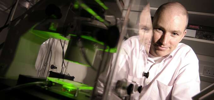
Back in 2015, Professor Stephen Taylor had a plan. After 15 years studying cell division in cell lines, he wanted to test how anti-mitotic chemotherapy drugs interfere with the process in a more clinically relevant setting.
Taylor and his team—based at the University of Manchester Division of Cancer Sciences—were keen to avoid some of the problems associated with cell lines. It's well documented that they often do not reflect the original tumor and few have any associated clinical information. So, they developed a plan to examine precisely how the microtubule stabilizer Taxol causes cancer cell death on patient-derived primary ovarian cancer cells.
As projects go, it was meant to be a relatively simple one. "I thought, right, let's actually get cells from patients," Taylor recalls. "Let's set up collaborations with a hospital and test our hypotheses on cells from biopsies."
Facilitated by the lab's location at The Christie hospital site in Manchester, it was a plan that, five years later, yielded a living biobank of ovarian cancer cells. The team can now collect samples—mainly in the form of ascites—from patients with chemo-naive and relapsed ovarian cancer. They have managed to get over 80 ex vivo ovarian cancer cultures growing.
While the plan was straightforward, the journey to this point proved to be anything but. "I was in for a massive surprise. What we uncovered was the dirty little secret of cancer biology, which everybody knows, but nobody tells," says Taylor. "Primary cancer cells do not grow at all well in culture."
Why a living biobank?
So, why go to the trouble? Ovarian cancer is the leading cause of gynecological-related mortality. High-grade serous ovarian carcinoma (HGSOC), the most prevalent sub-type, develops rapidly and is rarely diagnosed before advanced stage disease. Treatment options are limited and survival rates have not improved substantially for 20 years.
To develop new, more effective treatment, researchers need pre-clinical models that more closely reflect ovarian cancer in vivo. While cancer cell lines are an important resource, they come with a number of limitations. Perhaps most well-known is the simple fact that some well-established cell lines have existed for so long.
"Some have been growing in culture for 40, 50, maybe 60 years. If you think about genetic drift, and how those cells will have adapted—do they really reflect the original tumor from which they came?" asks Taylor. "We have very little knowledge about the origins of some of those cell lines. There's no pathology to go with them and there's certainly no clinical data."
It was the promise of a model system that more accurately reflected the phenotypic and genetic characteristics of ovarian cancer cells that spurred Taylor and his lab on to develop their living biobank. If they could collect and grow ovarian cancer cells directly from patients, they could avoid this genetic drift and annotate each culture with clinical information. Running on the assumption that the main barrier was access to patient samples, they set up a collaboration with The Christie and the Manchester Cancer Research Centre (MCRC) Biobank and began to collect the ascites of ovarian cancer patients.
"We'd solved the access problem. We now had samples coming out of our ears," says Taylor. "If we put them into a petri dish they should grow because established cell lines grow like weeds, right? That's when we were in for the massive surprise that primary cancer cells don't grow in culture."
Absolute magic
The problem seemed to be that ascites, while a good source of tumor cells, is also full of cancer-associated fibroblasts—which thrive in culture.
"What you see is the fibroblasts just growing and dividing, and eventually just taking over the entire dish," Taylor says. "Which made no sense. Cancer cells are immortal, whereas normal fibroblasts are mortal—so why were they growing like crazy and taking over my culture dish while the tumor cells just sat there doing nothing?"
Separated tumor cells didn't fare much better. They were convinced the answer to ovarian cancer cell proliferation was in the correct culture media conditions, but knowing the exact recipe was the sticking point. And it may have remained so were it not for a fortuitous comment during a CRUK program grant application review.
One of the reviewers pointed Taylor to a paper describing the decade-long search for the ideal culture medium to grow ex vivo human ovarian cancer cells. However, the process of getting hold of the medium was slow. In the meantime, Dr. Louisa Nelson, a postdoctoral research associate in the Taylor lab, read the paper and decided to take matters into her own hands.
"Louisa said 'why don't I just have a go?' And I just thought, no, there's no way that'll work—the recipe is so complicated," says Taylor.
But Nelson was determined, and it paid off—her medium worked and for the first time the Taylor lab could collect patient samples and then successfully grow ovarian tumor cells.

"When we got the first sample to grow, it was absolute magic," says Taylor. "To look down the microscope and see these little colonies of cells that were clearly growing. It was just amazing."
Despite her pivotal role, it's a success Nelson herself is humble about. "We now get a success rate of around 30% of patient samples establishing as a culture," she says. "This is still not ideal, but we are working to increase this by trying different culture conditions and different plastic coatings."
A path to patient avatars
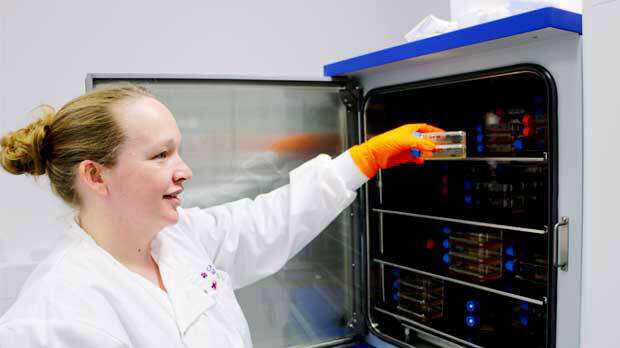
Nelson now runs the ovarian cancer models (OCMs) generated as part of the living biobank and has over 80 cultures growing—each with a clinical history. Key to this is the collaborations the lab has built.
"The MCRC Biobank plays a massive role in this," says Nelson. The MCRC is a partnership between CRUK, the University of Manchester and The Christie NHS Foundation Trust—and was set up to support collaborative research.
She adds: "They coordinate with the clinical staff at The Christie to enable us to receive the number of samples we do. They also coordinate consent from all the patients and provide us with treatment details. Without them the process wouldn't be as streamlined as it is."
Because the living biobank generates OCMs that so closely resemble the features of ovarian cancer, the Taylor lab can now use it to not only probe underlying biological mechanisms but also to test novel therapeutic strategies. Tantalizingly, the living biobank concept even opens the possibility of a personalized approach to ovarian cancer treatment—though this may be a way off yet.
"If we can rapidly grow cells in culture, it would be possible to test a range of drugs or drug combinations on each patient sample," Nelson explains. "This would then be fed back into the clinic, allowing clinicians to make informed decisions as to which drugs worked well for an individual patient. At the moment this isn't possible, but it's something we're working towards."
It's a cautious excitement shared by Taylor. "We want to take a patient biopsy and screen it against drugs and empirically determine what the patient might benefit from. In this way the OCM acts like a patient avatar. Without worrying about characterizing those cells or biomarkers…just let the tumor tell us what it is sensitive to."
"But there are a lot of challenges with this approach," adds Taylor. "There is no guarantee we can generate a culture, and it can take quite a bit of time. We haven't optimized the platform to do this yet."
For now, using the biobank to build an understanding of underlying biology has already yielded a surprise. Analysis of centrosome numbers and high-resolution time-lapse microscopy revealed far more pronounced heterogeneous mitoses than observed in established cell lines. This finding has led to a paper and suggests, says Taylor, that mitotic dysfunction in advanced human cancers is underestimated.
"When I first saw images of these cancer cells down the microscope they just looked so abnormal," he says. "You could just tell straight away that something was really horribly wrong. We're studying these cells as fresh out of a patient and what we get out is very chromosomally unstable. Yet the cells were surviving."
A lot of Taylor's samples are from patients who have undergone extensive chemotherapy with tumors that have acquired drug resistance. "The longer we leave the cells in culture they will become much more stable, much more homogeneous," he says. "As they evolve, some of that chromosomal damage will be lost. By trying to study them as soon as we can, I think we're overcoming a lot of the problems we see with cell lines."
Breadth and depth
So, what do the team want to do next with the living biobank? It's a matter of both breadth and depth says Taylor.
The breadth is simple—he wants to create as many OCMs from as many samples as possible. "Then we can start looking at any given drug and screen it on all our samples," says Taylor.
As for depth, that'll see the team focus on the specific details of a single sample. Because of the efficiency of the flow of ascites samples from the clinic to the lab, Taylor and Nelson can collect longitudinal sample sets from single patients. They can then begin to build a picture of how the tumor cells change over the course of the disease.
"As a cell biologist what I want is primary tumor cells prior to chemotherapy, and I want the relapsed, therapy treated disease to see what's changing," explains Taylor.
Finally, Taylor is very keen that the living biobank becomes a platform for collaborations.
"We can't do everything and there are lots of people who can make progress in areas of cancer cell biology we're not experts in," he says. "Whether the biobank becomes a resource for others, or we help with making up the culture media, this is a great opportunity to reach out and form collaborations."


