This article has been reviewed according to Science X's editorial process and policies. Editors have highlighted the following attributes while ensuring the content's credibility:
fact-checked
peer-reviewed publication
proofread
New insights into how sensory cells and neurons code for sound in our ears

Sensory cells and neurons in the ear communicate by secreting neurotransmitters in response to sound stimuli. Scientists of the University Medical Center Göttingen, the Cluster of Excellence Multiscale Bioimaging, and the Max Planck Institute for Multidisciplinary Sciences describe new details of this process that regulates the release of neurotransmitters and thus controls the transmission of sound stimuli. The results of this work were published in the Proceedings of the National Academy of Sciences.
Our nervous system contains about 100 billion nerve cells that communicate with each other at around 100 trillion contact points, known as synapses. Communication takes place via messenger substances that allow the transmission of information between sensory cells and neurons. These messenger substances allow us to process environmental stimuli, learn, and control our behavior.
They also play a fundamental role in the transmission of sound information in the process of hearing and can be the cause of hearing disorders. Such disorders are very common: according to the World Health Organization (WHO), about 466 million people, corresponding to around 5% of the world's population, suffer from disabling hearing loss. Understanding the elementary processes of hearing is an important prerequisite for developing better methods to treat hearing loss in the future.
Scientist Dr. Lina María Jaime Tobón and department director Prof. Dr. Tobias Moser, both working at the Institute of Auditory Neuroscience of the University Medical Center Göttingen (UMG), at the Cluster of Excellence "Multiscale Bioimaging: from Molecular Machines to Networks of Excitable Cells" (MBExC), and at the Max Planck Institute for Multidisciplinary Sciences (MPI-NAT), have now investigated the synapses between the sensory hair cells in the inner ear and the neurons of the auditory nerve.
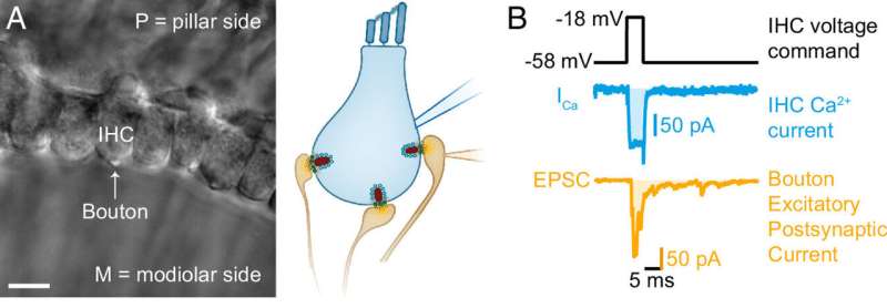
At these synapses, the incoming sound information is converted into a neuronal signal that accurately transmits the perceived sound to the brain. For the first time, individual synapses in the inner ear of hearing mice were analyzed using the patch-clamp technique that was pioneered by Erwin Neher and Bert Sakmann in Göttingen. This method makes it possible to "observe" the process of sound encoding.
The main focus of the scientists' work was on the question of how the synapses couple the release of the neurotransmitter, glutamate, to the sound stimulus. Glutamate is involved, among other things, in the transmission of stimuli between sensory and nerve cells.
Within the sensory cells, glutamate is transported to the synapse in "vesicles." The incoming sound activates calcium channels at the synapses, through which calcium ions enter the cell and trigger the release of glutamate. The results show how the release of glutamate increases with the strength of the stimulus, i.e., how a "sound signal" is converted into a "glutamate signal" and what role the calcium channels play in this conversion. The main actors in this process are calcium channels, calcium ions and synaptic vesicles, which are apparently a few millionths of a millimeter away from the channels.
Research results in detail
In hearing, sound waves are transmitted from the outer ear to the eardrum, which causes the small auditory ossicles in the middle ear to vibrate. These vibrations ultimately stimulate the sensory hair cells in the cochlea of the inner ear. The hair cells convert the mechanical stimulus into electrical activity and transmit this to the auditory nerve. The change in electrical potential of the cell is stronger in response to louder sounds.
At the synapse, the change in potential activates calcium channels, which subsequently allow calcium ions to enter the cell. Messenger substances, such as glutamate are transported in vesicles to the synapse. The calcium ions entering the cell through calcium channels in turn cause release of glutamate from the vesicles into the gap between the hair cell and the nerve cell. The glutamate can now activate the opposing nerve fibers of the auditory nerve, which transmit the sound information as nerve impulses to the brain.
In their study, the Göttingen scientists investigated the question of how the electrical discharge of the cell activates individual synapses to release neurotransmitters. The results show that a calcium channel and a vesicle always form a functional unit. Each of the calcium sensors located on the vesicles, which ensure the release of messenger substances, must apparently bind four calcium ions before messenger substances are finally sent to the neighboring nerve cell of the auditory nerve.
"For the first time, we were able to investigate the coupling of calcium channels and neurotransmitter release at individual hair cell synapses with the highest time resolution. This way we could study the millisecond-short first phase transmitter release in great detail," says Dr. Tobón, who is also a member of the MBExC's Hertha Sponer College. "This has enabled us to reliably determine the number of calcium ions that need to bind to the synaptic vesicle sensor for release."
"This breakthrough is an exciting example of the work in the MBExC Cluster of Excellence," says Prof Dr. Moser, senior author of the publication. "Our previous work had already provided initial indications of a close spatial coupling of the glutamate release from a synaptic vesicle to the calcium influx through neighboring calcium channels. The high sensitivity of the measurements carried out here now offers the most precise quantification of this process known to us, which takes place on a scale of a few millionths of a millimeter," says Moser.
More information: Lina María Jaime Tobón et al, Ca 2+ regulation of glutamate release from inner hair cells of hearing mice, Proceedings of the National Academy of Sciences (2023). DOI: 10.1073/pnas.2311539120


