This article has been reviewed according to Science X's editorial process and policies. Editors have highlighted the following attributes while ensuring the content's credibility:
fact-checked
trusted source
proofread
Q&A: Stem-immunity hubs associated with response to immunotherapy

Jonathan Chen, MD, Ph.D., an investigator in the Department of Pathology at Massachusetts General Hospital, and Nir Hacohen, Ph.D., director of the Center of Cancer Immunology at Massachusetts General Hospital, are co-authors of a recently published study in Nature Immunology, Human Lung Cancer Harbors Spatially-organized Stem-immunity Hubs Associated with Response to Immunotherapy.
Here, they discuss their findings.
What question were you investigating?
Multicellular networks are critical in mediating immune responses. How do immune cells organize within tumors to effectively eliminate malignant cells?
We recently reported the discovery of a network of immune cells found in colorectal cancer. We termed these networks "immunity hubs," and they are characterized as foci of activated T cells abutting tumors and immune myeloid cells expressing T cell-attracting molecules.
This finding suggests the existence of a positive feedback loop in which activated T cells drive further T cell recruitment by local cells in the tumor.
We reasoned that immunity hubs might be predictive of response to immunotherapy because they were enriched in a class of colorectal tumors known to have a higher rate of response to PD-1 blockade immunotherapy.
However, while our study of colorectal samples includes this class of tumors, the patients in our study were not treated with immunotherapy.
In this new study, we sought to answer whether immunity hubs are indeed predictive of response to standard-of-care immunotherapy.
We studied non-small cell lung cancer (NSCLC), which is commonly treated with PD-1 blockade immunotherapy and is the leading cause of cancer death worldwide.
What methods or approach did you use?
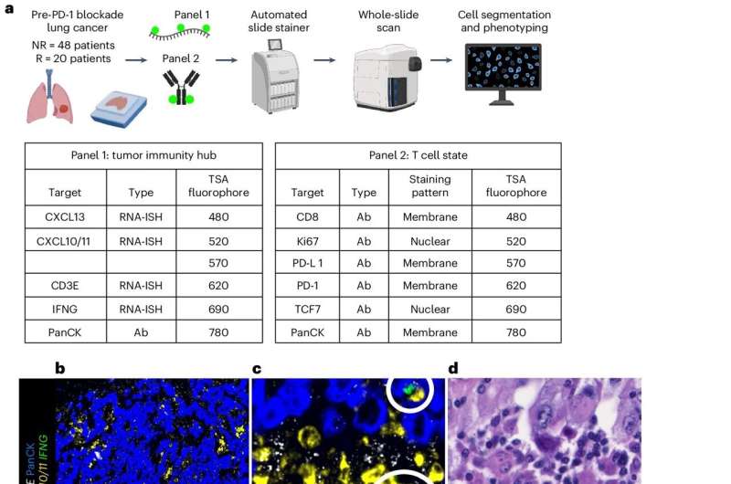
We first used a focused approach to image key transcripts and proteins of the hubs to detect immunity hubs in a cohort of 68 lung cancer patients, allowing us to discover a new variant of the immunity hub, which we termed the "stem-immunity hub."
We then developed a less biased approach to image 479 transcripts that represent many cell types and gene programs in the tumor microenvironment to better understand the composition of these hubs and their interactions with other cells in the tumor.
This high-resolution spatial approach allowed us to discover important cell-cell interactions that may regulate the formation and function of these hubs.
What did you find?
We found that patients without immunity hubs in their tumors prior to PD-1 blockade therapy had poor outcomes relative to patients with hubs.
Critically, we discovered the stem-immunity hub, a subtype of immunity hub that is strongly associated with favorable immunotherapy response.
Stem-immunity hubs were enriched for a type of T cell remarkable for its ability to divide and invigorate the antitumor immune response after PD-1 blockade. So, the finding of these stem-immunity hubs represents a new way to think about how the immune response is organized in human cancer.
What are the implications?
The most obvious implication of our work is that we can use the presence of the immunity hub, particularly the stem-immunity hub subclass, as a biomarker to predict response to immunotherapy.
The current standard biomarker for prediction of immunotherapy response is PD-L1 antibody staining, but it can be inaccurate and difficult to use.
We are currently testing a simple two marker tissue stain that reflects immunity and can be assessed by pathologists using a standard workflow.
We think the immunity hub may be how the immune system organizes to fight tumors.
Therefore, we aim to develop therapeutics based on findings in this study to augment immunity hubs and support the anti-tumor immune response.
What are the next steps?
As stated above, we are testing a simple two-color slide stain for stem-immunity hubs that may be clinically useful for guiding treatment decisions.
Additionally, we hypothesize that stem-immunity hubs may be sites where stem-like T cells are nurtured and could serve as platforms for launching a durable anti-tumor response.
We are currently testing the role of stem-immunity hubs in mouse models to determine if they induce greater and more durable anti-tumor immune response.
More information: Jonathan H. Chen et al, Human lung cancer harbors spatially organized stem-immunity hubs associated with response to immunotherapy, Nature Immunology (2024). DOI: 10.1038/s41590-024-01792-2


