This article has been reviewed according to Science X's editorial process and policies. Editors have highlighted the following attributes while ensuring the content's credibility:
fact-checked
trusted source
proofread
AI model may yield better outcomes for prostate cancer

Investigators from UCLA Health found using artificial intelligence to help map out the boundaries of cancerous prostate tissue can significantly reduce the risk of underestimating the extent of prostate cancer—an advancement that can help ensure an accurate diagnosis, precise treatment planning and effective surgical procedures.
By using AI to assist with cancer contouring, the researchers found predicting the cancer size was 45 times more accurate and consistent than when physicians used only conventional clinical imaging and blood tests to predict the cancer extent.
The findings were published in the Journal of Urology.
"Accurately determining the extent of prostate cancer is crucial for treatment planning, as different stages may require different approaches such as active surveillance, surgery, focal therapy, radiation therapy, hormone therapy, chemotherapy, or a combination of these treatments," said study author Dr. Wayne Brisbane, assistant professor of urology at the David Geffen School of Medicine at UCLA and member of the UCLA Health Jonsson Comprehensive Cancer Center.
Assessing the extent of prostate cancer is a complex task and typically requires a surgeon to consider various diagnostic tests such as a prostate-specific antigen (PSA) blood test, imaging tests like MRI, CT scans, and other clinical features simultaneously to determine the aggressiveness of the cancer cells.
Physicians tend to rely on a tumor's MRI appearance, but the true extent of the prostate cancer can be "MRI-invisible" causing physicians to underestimate the tumor size, noted Brisbane. AI could help resolve this challenging problem.
The new AI system, developed by researchers at UCLA and Avenda Health, has already shown to better define the margins of prostate cancer than MRI, demonstrating the potential of AI to help improve minimally invasive treatment approaches like focal therapy, which is a relatively new approach for treating prostate cancer that aims to eliminate the cancer cells while minimizing damage to surrounding healthy tissue. However, prior to this study, characterizing the performance of the AI system in the hands of physicians was previously not tested.
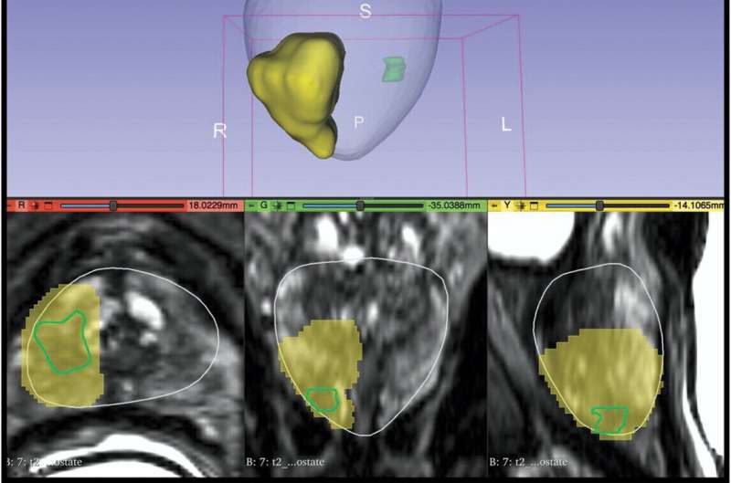
In order to evaluate the cancer contouring and clinical decision-making of physicians with and without AI software, the researchers conducted a multi-reader-multi-case study that compared cognitive and hemi-gland contouring methodologies to AI-assisted contours.
Seven urologists and three radiologists from different hospitals with varying levels of experience ranging from two to 23 years reviewed cases of 50 patients who had undergone a prostatectomy but who might have been eligible for focal therapy.
Each case included images from a specific type of MRI scan called T2-weighted MRI, along with outlines of the prostate gland and areas where cancer was suspected, as well as a report from a biopsy.
First, the physicians looked at the images and manually drew outlines around the suspected cancerous areas, aiming to encapsulate all significant disease. Then, after waiting for at least four weeks, they reexamined the same cases, this time using AI software to assist them in identifying the cancerous areas.
An analysis was then completed to evaluate the accuracy and negative margin rate—which indicates whether all cancerous tissue was identified—of the cancer outlines drawn by each method.
The researchers found when using conventional means, doctors only achieved a negative margin 1.6% of the time. When assisted by AI the number increased to 72.8%.
"We saw the use of AI assistance made doctors both more accurate and more consistent, meaning doctors tended to agree more when using AI assistance," said Shyam Natarajan, assistant adjunct professor of urology, surgery, and bioengineering and senior author of the study.
The team also found that the use of AI increased clinician recommendations for focal therapy among patients with unilateral cancer and reduced variation in accurate tumor encapsulation, which could help reduce the risk of side effects commonly associated with more aggressive treatments like surgery or radiation therapy.
"Overall, the use of AI in cancer treatment could lead to more effective and personalized care for patients, with treatments that are better tailored to their individual needs and more successful in fighting the disease," said Brisbane.
The study's co-first authors are Sakina Mohammed Mota and Alan Priester, from Avenda Health. Other authors include James Sayre from UCLA and Joshua Shubert, Jeremey Bong and Brittany Berry-Pusey from Avenda Health.
Conflicts of Interest: Mota, Priester, Shubert, and Bong are employees of Avenda Health. Berry-Pusey and Natarajan are cofounders of Avenda Health. Sayre consults for Avenda Health.
More information: Sakina Mohammed Mota et al, Artificial Intelligence Improves the Ability of Physicians to Identify Prostate Cancer Extent, Journal of Urology (2024). DOI: 10.1097/JU.0000000000003960


