Scientists reveal endocardial origin of liver vasculature

On March 29, Nature Genetics published a research article titled "Genetic lineage tracing identifies endocardial origin of liver vasculature," from Prof. ZHOU Bin's lab at the Institute for Nutritional Sciences (INS), Shanghai Institutes for Biological Sciences, CAS.
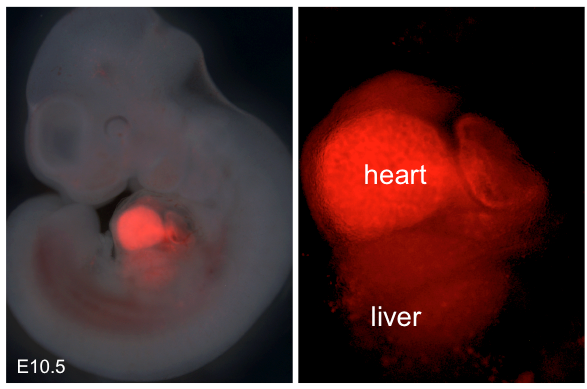
Taking advantage of genetic lineage tracing and tissue-specific gene knockout technology, researchers found that part of the liver vasculature is derived from the endocardium in the developing heart. During this process, endocardial VEGF/VEGFR2 signaling plays an important role in liver angiogenesis and organogenesis.
Blood vessels are essential for liver function during homeostasis and regeneration after injury. During embryonic development, blood vessels form passive conduits to deliver oxygen and nutrients. Researchers establish instructive development niches to promote liver organogenesis before the onset of circulation. Determining the developmental program of hepatic vessel formation provides the foundation for the design of strategies to stimulate vessel growth upon liver regeneration.
Taking advantage of genetic mouse tools that specifically label endocardium, researchers carried out lineage tracing experiments and found that endocardial cells in the early embryonic heart could migrate and form a primitive vascular plexus surrounding the liver bud.
With development of the liver, this vascular plexus, which plays important roles in liver organogenesis, differentiates into central veins, sinusoid endothelial cells, portal veins and arteries. Interestingly, these endocardium-derived endothelial cells could also form part of the lymphatic endothelial cells. Notably, these endocardium-derived liver vessels respond to injury and proliferate to form new vessels during liver regeneration after partial hepatectomy.
By intersectional lineage tracing technology, researchers pinpointed that endocardium-derived liver vasculature is derived from sinus venosus endocardium, which is also the origin of coronary vessels. This is the first study to demonstrate that there exists a common origin for liver vasculature and coronary vessels—sinus venosus.
The VEGF/VEGFR signaling pathway, which is essential for angiogenesis, plays important roles in proliferation, migration, survival and penetration of vascular endothelial cells. There are three members in the VEGFR family: VEGFR1 (Flt1), VEGFR2 (KDR/Flk1), and VEGFR3 (Flt4). VEGFR2, which is activated through binding to VEGF, is mostly distributed in vascular endothelial cells. In this study, researchers found high expression of VEGF in hepatocytes during liver bud development, and high expression of VEGFR2 in the endocardium.
This expression map indicated that there might exist interactions between hepatocytes and the endocardium through the VEGF/VEGFR2 signaling pathway. Tissue-specific deletion of VEGFR2 in the endocardium led to a significantly reduced amount of endocardium-derived vessels in livers, and dysplastic livers only one-quarter of the size compared of controls. Thus, the VEGF/VEGFR2 signaling pathway may affect liver organogenesis through regulating formation of endocardium-derived liver vessels.
More information: Hui Zhang et al. Genetic lineage tracing identifies endocardial origin of liver vasculature, Nature Genetics (2016). DOI: 10.1038/ng.3536


